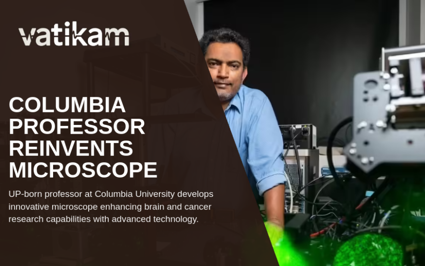
Revolutionizing Medical Imaging: Meerut-Born Professor Reinvents Microscope Technology at Columbia University
Medical imaging plays a pivotal role in understanding complex biological tissues such as the human brain and cancer tumors. Recently, a breakthrough in microscope technology promises to transform this field, making detailed three-dimensional (3D) imaging more accessible and affordable. This revolution is led by Raju Tomer, a biology professor born in Meerut, working at Columbia University.
The Challenge in Medical Imaging
Traditional microscopes used for tissue imaging have faced a challenging compromise: either they provide sharp, high-resolution images but lack the ability to penetrate deeply into tissues, or they can image deeper but at the cost of clarity. This limitation is largely due to the technical differences between expensive oil-immersion lenses and cheaper air lenses.
Oil-immersion lenses, while yielding crystal-clear images, struggle to scan deep tissues, whereas air lenses can penetrate deeper but produce blurry images, especially when used with chemicals that make tissues transparent for 3D viewing. Researchers have long sought a solution that balances depth penetration and image quality without the burden of cost and complexity.
Introducing HySIL: A Breakthrough Optical System
Addressing this challenge, Professor Raju Tomer and his team have developed HySIL (Hybrid Solid-Liquid Optics). This innovative system ingeniously combines a curved solid lens with a specially matched immersion liquid, functioning as a continuous optical system. The HySIL design allows simple, inexpensive air lenses to achieve image sharpness comparable to costly lab equipment while maintaining the ability to image deep within tissues.
According to Tomer, the system works like a perfect puzzle where the solid lens and liquid fit seamlessly, enabling detailed and sharp 3D imaging across various tissue samples and preparation methods. This advancement drastically reduces microscope complexity and cost, making advanced imaging more accessible to laboratories worldwide.
Applications of HySIL Technology
Tomer’s team has transformed this concept into practical tools, including a compact add-on device compatible with existing microscopes globally. They also created a high-resolution variant known as the “Super-Scope” and a small commercial microscope called “Slice,” developed in 2024.
Using these technologies, researchers have successfully imaged entire mouse, salamander, and cavefish brains, lab-grown miniature human brain tissues, as well as real human cancer biopsies in full 3D. This comprehensive imaging capability represents a significant leap beyond the traditional practice of examining tissue slices on glass slides, providing a holistic view of tissue architecture.
The Impact on Brain and Cancer Research
Traditionally, medical professionals and researchers have relied on viewing thin, two-dimensional tissue slices, which limits observation to a single plane and often misses the broader tissue structure. The ability to visualize tissue in 3D allows an unprecedented understanding of biological details and their spatial relationships.
Pathology experts such as Hanina Hibshoosh from Columbia University Irving Medical Center emphasize that affordable 3D imaging tools like HySIL will be increasingly essential. With advancements in artificial intelligence (AI) enabling analysis of vast tissue data sets, high-quality, deep-tissue imaging becomes critical to improved diagnosis, prognosis, and biomedical research.
Conclusion
Professor Raju Tomer’s groundbreaking invention of the HySIL microscope system represents a transformative advance in medical imaging technology. By combining affordability, simplicity, and high-resolution 3D imaging capability, HySIL stands to enhance brain and cancer research significantly. This innovation is not only a testament to scientific creativity but also a promising beacon for future breakthroughs in medical diagnostics and research.